4. Human body model architecture
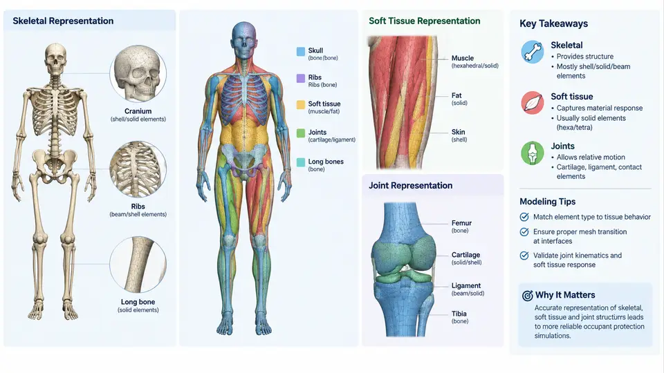
A finite element (FE) human body model (HBM) is not a single mesh but a layered, multi-material assembly whose architecture determines what physical responses the model can predict. In occupant protection, the three structural pillars are the skeleton, the soft tissue, and the joints that connect skeletal segments. Each pillar carries a distinct mechanical role, uses different element formulations and material laws, and contributes differently to injury metrics such as bone fracture, organ strain, and joint kinematics. Understanding this architecture is a prerequisite for selecting the right model for a given crash scenario and for interpreting its output correctly.
Skeletal Representation: Rigid Bodies Versus Deformable Bone
The skeleton provides the primary load path and defines the overall geometry of the occupant. In FE HBMs, skeletal segments are represented in one of two ways. Rigid body representations treat a bone as a non-deforming body with mass and inertia, connected to neighbors through joint elements. They are computationally cheap and are common in simplified models used for gross kinematics studies. Deformable bone representations use shell or solid elements with elastic–plastic material laws so that stress and strain can be resolved locally, enabling fracture prediction.
Cortical bone is typically modeled with shell elements assigned an elastic–plastic material (for example, a Young's modulus on the order of 15–20 GPa and a yield stress around 100–150 MPa for long bones, values that vary by bone and loading rate). Trabecular bone, where present, is modeled with lower-modulus solid elements. The choice of element size matters: a rib cage meshed at 5 mm average shell edge length can capture rib fracture patterns under belt loading, whereas a 15 mm mesh may smear out stress concentrations and under-predict fracture. A practical rule is that the smallest anatomical feature of interest should be spanned by at least two to three elements.
Soft Tissue Representation: Organs, Fat, Muscle, and Skin
Soft tissue governs energy absorption, organ deformation, and the coupling between the skeleton and the environment (belt, airbag, seat). It is the most material-sensitive part of an HBM. Typical representations include:
- Solid elements for organs such as the brain, liver, spleen, and heart, using hyperelastic or viscoelastic material laws to capture rate-dependent stiffness.
- Solid or thick-shell elements for muscle and fat layers, often modeled with low-modulus, nearly incompressible materials.
- Membrane or shell elements for skin and fascia, providing surface continuity and contact definition.
Because soft tissue is nearly incompressible, element formulations must handle a Poisson's ratio close to 0.5. Reduced-integration solid elements with hourglass control, or selectively reduced integration, are standard choices. A common failure mode in soft tissue modeling is hourglassing, where under-integrated elements deform in spurious zero-energy modes; this appears as a jagged, checkerboard-like deformation pattern in the organ and is corrected by increasing hourglass stiffness or refining the mesh.
Material parameters for soft tissue are usually derived from cadaveric or animal testing and are strain-rate dependent. For example, brain tissue is often modeled with a viscoelastic shear modulus that relaxes over tens of milliseconds, which is why head impact duration strongly influences predicted strain. When porting parameters between models, always confirm the unit system and the reference strain rate, since a modulus quoted at quasi-static rates will under-predict stiffness in a 10–20 ms impact.
Joint Representation: Kinematics and Load Transfer
Joints are the interface between skeletal segments and are where the model's kinematic fidelity is largely decided. Three common approaches exist:
| Joint type | Typical implementation | Best suited for |
|---|---|---|
| Kinematic (constrained) | Rigid links, revolute or spherical constraints | Gross motion, simplified models |
| Deformable (ligamentous) | Springs, beams, or ligament elements with nonlinear force–displacement curves | Joint injury, ligament rupture, realistic range of motion |
| Contact-based | Articular surfaces with contact definition and cartilage layers | Detailed joint mechanics, cartilage stress |
Ligamentous joints are the most common compromise in full-body HBMs. Each ligament is represented by one or more nonlinear spring or beam elements whose force–elongation curve is calibrated to experimental data. The knee, for instance, uses separate elements for the anterior cruciate ligament (ACL), posterior cruciate ligament (PCL), medial and lateral collateral ligaments, each with distinct stiffness and failure elongation. When a ligament element reaches its failure strain, it is deleted or its stiffness drops to near zero, allowing the joint to dislocate — a behavior that directly affects lower-extremity injury prediction in frontal impacts.
How the Three Layers Interact
The architectural challenge is that skeleton, soft tissue, and joints are not independent. Soft tissue mass loads the skeleton; skeletal motion drives soft tissue deformation; joints transmit and redistribute loads between segments. In a frontal sled simulation, the sequence is roughly: belt engages the chest, chest soft tissue compresses and loads the rib cage, ribs deflect and transfer load through the thoracic spine joints, and the head–neck complex responds through cervical joint stiffness. If any layer is misrepresented, the error propagates.
A useful verification practice is to isolate each layer before running a full-body case. Prescribe a known displacement to a single bone and check that the joint rotates within its physiological range; apply a known pressure to an organ and compare the resulting strain against published tissue test data; load a rib cage in isolation and compare force–deflection response to cadaveric corridors. Only after each layer passes should the assembled model be used for injury assessment.
Practical Implications for Model Selection and Use
The architecture directly constrains what a model can answer. A model with rigid bones and kinematic joints cannot predict rib fracture or ligament rupture, regardless of how refined its soft tissue mesh is. Conversely, a fully deformable model with detailed ligaments is computationally expensive and may be unnecessary for a study focused on head kinematics. The practical workflow is to match the model's architectural fidelity to the injury mechanism under investigation, then verify each layer independently before trusting the assembled response.
When reviewing an HBM's documentation, check three things: the element type and material law for each tissue, the source and calibration range of material parameters, and the joint failure criteria. These three items determine whether the model's predictions are credible for your specific loading condition. A model that performs well in a frontal 40 km/h sled test may behave poorly in a lateral impact if its joint stiffness and soft tissue parameters were calibrated only for frontal loading.