1. Hip femur and knee injury diagnosis
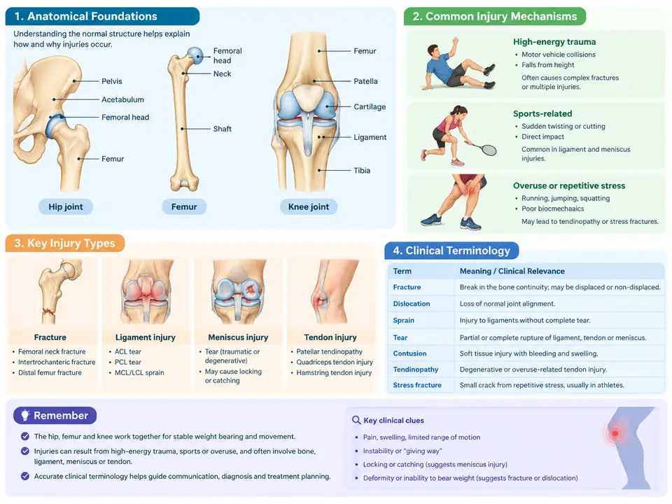
Effective diagnosis of hip, femoral, and knee injuries begins long before the first imaging study is ordered. The clinician must hold a precise mental model of the regional anatomy, understand how force is transmitted through bone and soft tissue during injury, and speak a shared vocabulary that allows findings to be communicated unambiguously across the care team. This chapter establishes that foundation: the bony and soft-tissue architecture of the hip, femur, and knee; the mechanical pathways along which injurious loads travel; and the standardized terminology used to describe location, mechanism, and severity. These three elements are interdependent — a term such as "varus collapse" is meaningless without an anatomical reference frame, and a mechanism such as "dashboard impact" only guides diagnosis when the clinician can trace the force path from patella to posterior acetabular wall.
Regional Anatomy as a Diagnostic Map
The hip joint is a ball-and-socket articulation between the femoral head and the acetabulum of the pelvis. Its stability derives from a deep socket, a strong fibrous capsule, and the acetabular labrum, which deepens the socket and seals the joint. The femoral head receives its blood supply predominantly from the medial circumflex femoral artery via the retinacular vessels; this vascular anatomy explains why displaced intracapsular femoral neck fractures carry a high risk of avascular necrosis, while extracapsular intertrochanteric fractures generally do not. The proximal femur is divided into the head, neck, greater and lesser trochanters, and the intertrochanteric and subtrochanteric regions — divisions that map directly onto fracture classification and treatment decisions.
The knee is a modified hinge joint comprising the tibiofemoral and patellofemoral articulations. The tibiofemoral joint relies on the anterior and posterior cruciate ligaments for anteroposterior and rotational constraint, and on the medial and lateral collateral ligaments for varus-valgus stability. The menisci distribute load and improve congruity; the patella acts as a sesamoid pulley that increases the extensor mechanism's moment arm. The distal femur and proximal tibia form the metaphyseal and epiphyseal regions where many fractures occur, and the physis in skeletally immature patients is a distinct weak point that shapes pediatric injury patterns.
Injury Mechanisms and Force Pathways
Injury mechanism describes how energy was delivered to the body. Three broad categories dominate hip, femur, and knee trauma: low-energy falls from standing height, high-energy impacts such as motor vehicle collisions or falls from height, and repetitive or overuse loading. The mechanism predicts both the fracture pattern and the associated soft-tissue injury, and it should be recorded in the patient's own words alongside the clinician's interpretation.
In a typical low-energy hip fracture, an elderly patient falls onto the greater trochanter. The impact generates an axial and bending load through the femoral neck, and the bone fails in compression on the inferior side and tension on the superior side. This produces the classic displaced transcervical or intertrochanteric pattern. In a high-energy dashboard injury, the flexed knee strikes the dashboard, driving the femoral shaft posteriorly and often producing a posterior hip dislocation or a distal femoral fracture; the same impact can rupture the posterior cruciate ligament. Valgus loading of the knee with a planted foot — common in sports — stresses the medial collateral ligament and may tear the medial meniscus, while a direct blow to the lateral knee can produce a lateral tibial plateau fracture.
Understanding the force path also guides the physical examination. A patient with a posterior hip dislocation typically presents with the limb shortened, adducted, and internally rotated, because the femoral head sits behind the acetabulum. An anterior dislocation presents with the limb externally rotated and abducted. These positional signs are direct consequences of the direction in which the femoral head displaced, and they should be checked before any manipulation.
Clinical Terminology and Its Diagnostic Role
Standardized terminology allows a finding to be communicated without ambiguity. Four descriptor families are essential in this region. First, location: anatomical region and side, using terms such as "intracapsular," "extracapsular," "basicervical," "subtrochanteric," "supracondylar," and "tibial plateau." Second, displacement and angulation: displacement describes translation of the distal fragment relative to the proximal one, while angulation describes the axis change. Varus angulation means the distal segment points toward the midline; valgus means it points away. Third, comminution and articular involvement: a fracture is comminuted when it has more than two fragments, and intra-articular when the fracture line enters the joint surface — a distinction that changes management because articular step-off must be reduced. Fourth, stability and neurovascular status: a fracture is unstable if it cannot maintain its reduction under physiological load, and the documentation must record distal pulses, capillary refill, and sensation, since a displaced supracondylar femoral fracture can injure the popliteal artery.
Several terms are frequently misused and deserve clarification. "Dislocation" means complete loss of articular contact; "subluxation" means partial loss. "Sprain" refers to ligament injury graded I to III by the degree of fiber disruption and laxity, whereas "strain" refers to muscle-tendon injury. "Open fracture" means the fracture communicates with the external environment through a skin wound — a designation that mandates urgent antibiotics and surgical debridement, not merely wound closure. Using these terms precisely is not pedantry; each one triggers a different clinical pathway.
Putting the Framework into Practice
Consider a worked example that integrates anatomy, mechanism, and terminology. A 72-year-old patient is brought in after a fall from standing. The limb is shortened and externally rotated. The clinician records: "Right hip pain after low-energy fall onto the greater trochanter; right lower limb shortened by approximately 2 cm and externally rotated; distal pulses intact; no open wound." The mechanism (low-energy axial and bending load) and the positional sign (shortening with external rotation) point to a displaced femoral neck or intertrochanteric fracture. The terminology distinguishes the two: an intracapsular femoral neck fracture raises avascular necrosis concern and may warrant arthroplasty in an elderly patient, while an extracapsular intertrochanteric fracture is typically treated with internal fixation. The documented neurovascular status establishes a baseline for later comparison.
For a second example, a 24-year-old athlete plants the foot and pivots, feeling a pop on the medial knee. The clinician records: "Valgus load with planted foot; medial joint line tenderness; valgus stress test opens the medial compartment at 30° of flexion; no locking." The mechanism and the positive valgus stress test localize the injury to the medial collateral ligament, and the absence of locking argues against a displaced meniscal tear. Grading the sprain (I–III) and documenting the opening in degrees provides an objective baseline for follow-up.
Key Takeaways
- Anatomical naming — head, neck, trochanters, condyles, plateau, and the ligamentous and vascular structures — is the reference frame for every diagnosis in this region.
- Injury mechanism predicts the fracture pattern and the associated soft-tissue injury; record both the patient's account and the clinician's interpretation.
- Use standardized descriptors for location, displacement and angulation, comminution and articular involvement, and stability and neurovascular status.
- Distinguish dislocation from subluxation, sprain from strain, and open from closed fractures, because each term changes the clinical pathway.
- Document distal pulses, sensation, and limb position before and after any intervention to establish a defensible baseline.